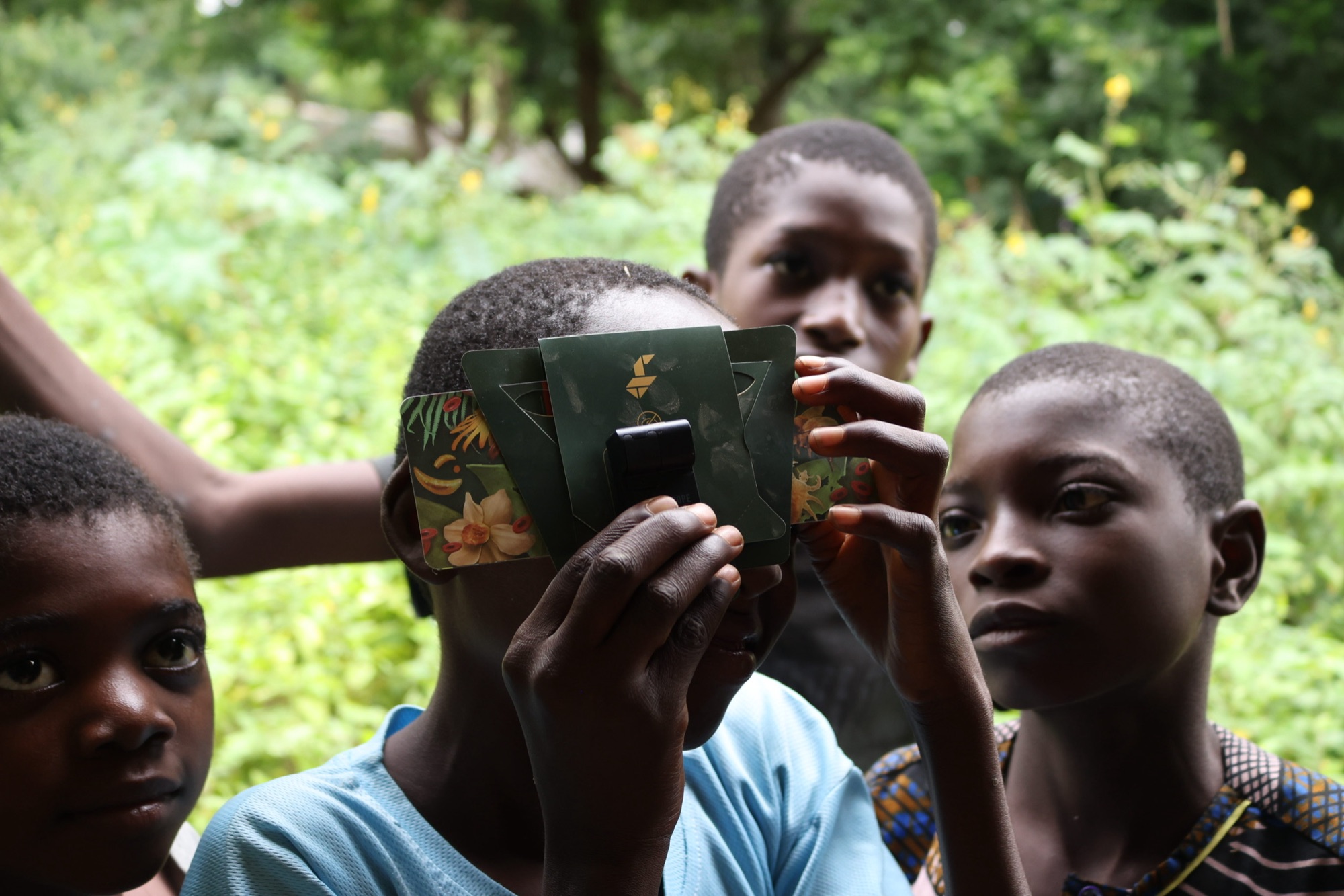
A slit lamp is one of the most basic instruments in an eye exam. It’s also expensive, bulky, and — for most of the world — only ever found inside a well-resourced clinic. I’m interested in a practical workaround.
Picture the standard tool: a binocular microscope and a precisely shaped beam of light that lets a clinician examine the front of the eye — the cornea, iris, and lens — in fine detail. It’s indispensable for diagnosing cataracts, uveitis, corneal injuries, and more. But the equipment carries a clinic-grade price tag and a clinic-sized footprint, which means the exam simply doesn’t travel to the people who can’t travel to it.
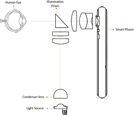
The workaround I keep coming back to: a portable, 3D-printed slit-lamp attachment paired with a smartphone, so a clinic — or a screening table in a community hall — can capture high-resolution anterior segment images without a full traditional unit.
It already mostly works
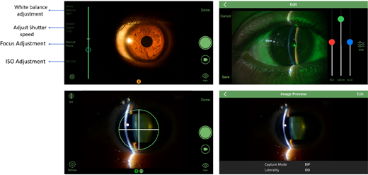
This isn’t wishful thinking. A recent paper describes a handheld system built from exactly these parts: a warm-white LED light pen housed in a custom case, a biconvex lens that focuses the light through a narrow 0.4 mm slit, and a smartphone fitted with a 100 mm macro lens serving as the camera. Simple, printable, cheap.
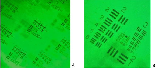
In a small head-to-head study, thirteen patients were examined with both the portable rig and a conventional slit lamp, producing twenty-six images. Three ophthalmologists — each with at least three years of experience — then rated the images blind, on image quality, diagnostic usefulness, and ease of use.
How the printable rig scored
Specialists rated the images comparable to conventional slit-lamp photos, and rated the device itself as easy to use. Source: Caruso et al., European Journal of Ophthalmology, 2025.
Comparable image quality, a fraction of the cost, and it fits in a backpack.
From a paper to a clinic in 2026
The idea for 2026 is straightforward: take an existing low-cost design like this one and validate it in a real clinic workflow — in Houston, or another site where we have access to low-cost clinics. The aim isn’t to invent new optics. It’s to prove the workflow holds up with real staff, real patients, and real time pressure.
What the pilot has to answer
Does the image hold up where it matters?
Focus on conditions where an anterior photo genuinely changes care — cataracts, uveitis — and compare results directly against a standard slit-lamp exam.
How heavy is the training burden?
Train staff on a simple capture protocol, then measure how quickly they get usable images. A tool only counts if a busy clinic can actually adopt it.
Do clinicians trust it?
Gather quick feedback through short surveys — on image quality, confidence, and whether they’d reach for it again.
If it performs well, the destination is a simple, ready-to-use kit: the printed attachment, a straightforward training checklist, and an image-capture guide — something any clinic can pick up and use without waiting on high-cost equipment. That’s the whole thesis of the Idea Garden in one object: take a proven low-cost design, and do the unglamorous work of making it usable.
Sources & further reading
- Caruso A, et al. A 3D-printed portable slit lamp for high-resolution anterior segment images. European Journal of Ophthalmology, 2025.doi.org ↗
- Dutt S, et al. Design and Performance Characterization of a Novel, Smartphone-Based, Portable Digital Slit Lamp. Translational Vision Science & Technology, 2021.doi.org ↗
An early idea, looking for a clinic.
This one is still a concept note — but a fundable, buildable one. If you run or know a low-cost clinic in Houston that sees anterior-segment cases, or you want to help print and test the first prototype, we’d love to hear from you.