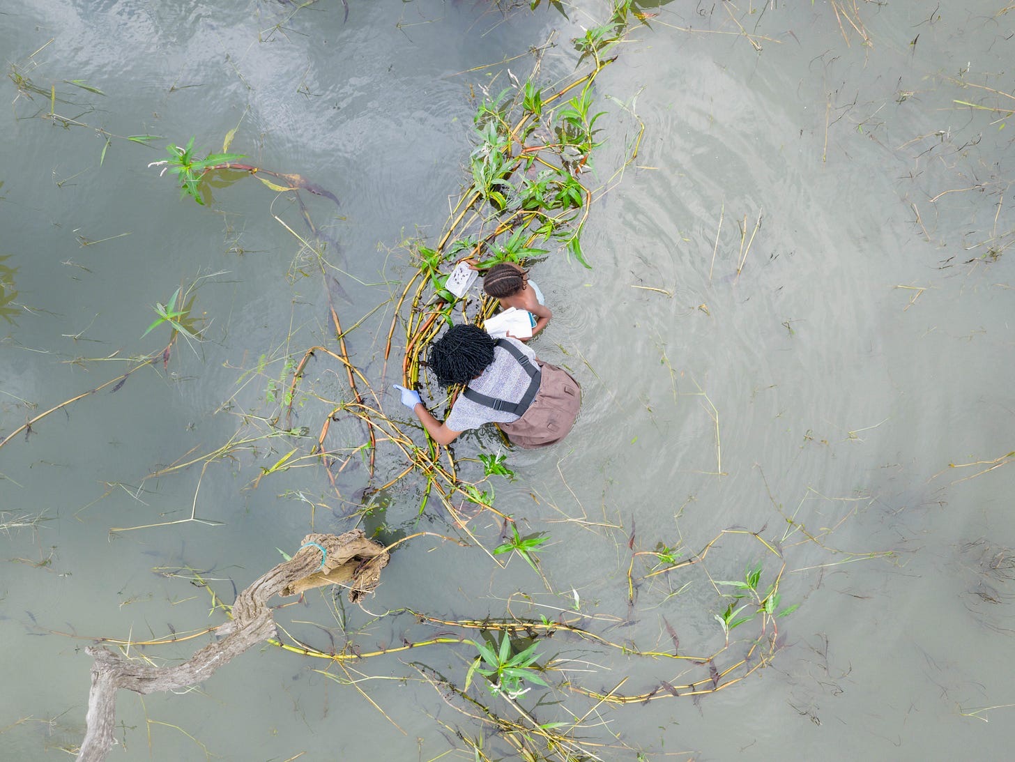
 UCSF · Graham Lab
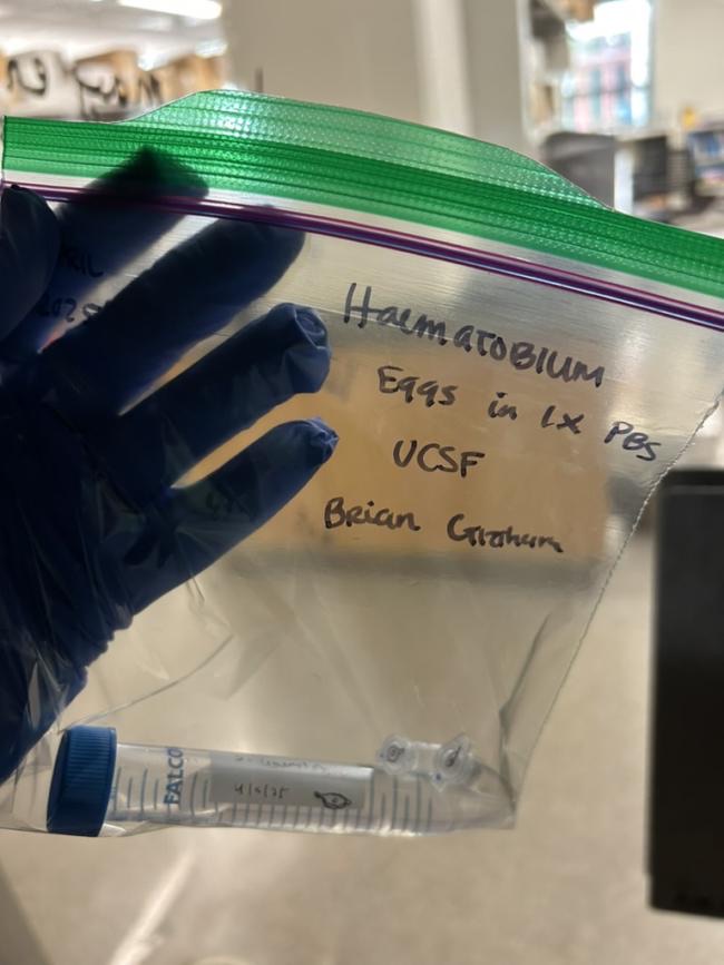
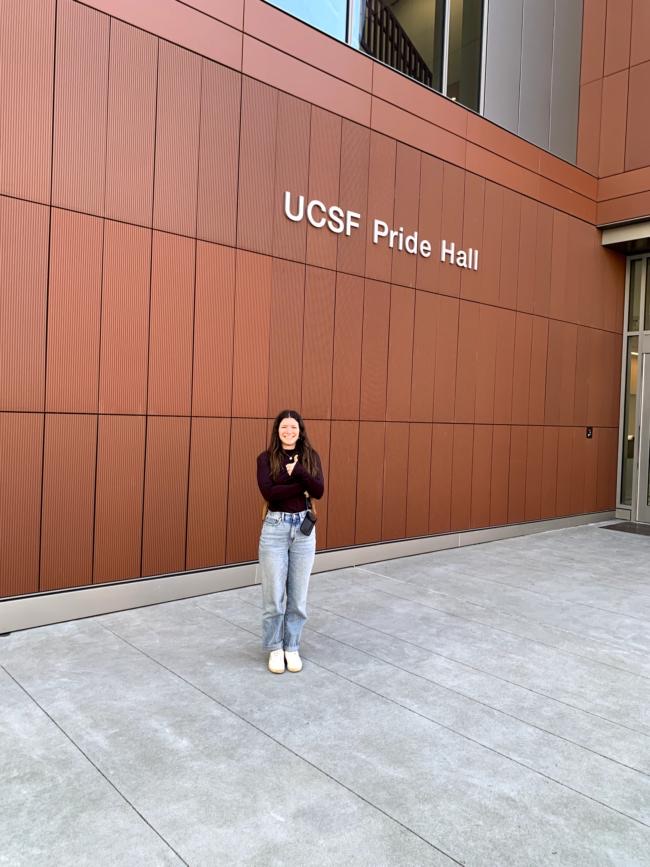
 Sometimes the most extraordinary samples come from the most unexpected connections. Through a friend of a friend, we found the lab of Dr. Brian Graham at UCSF — who handed our team one of the stranger gifts in global health research: a vial of Schistosoma haematobium eggs.
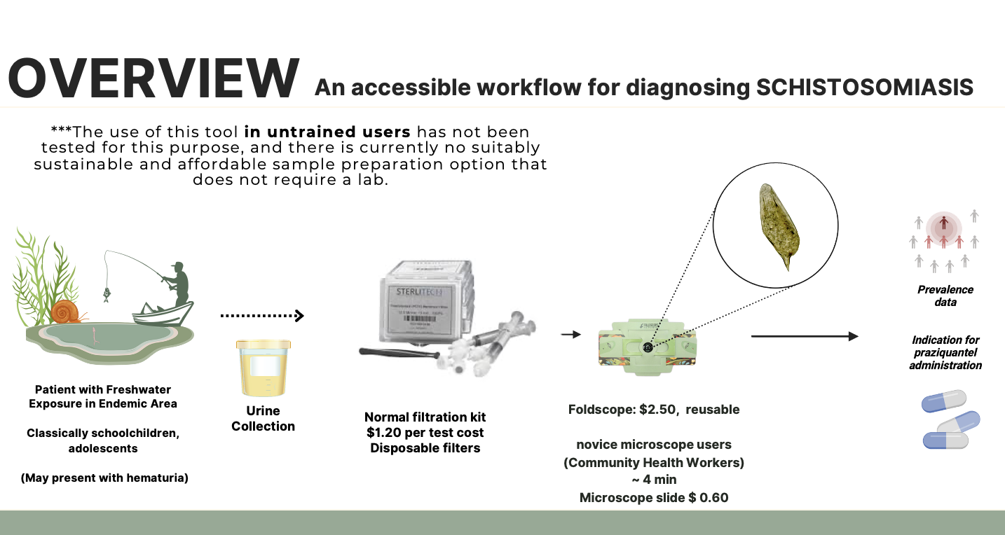
This is the story of the S. haematobium eggs we collected at UCSF, carried to another Bay Area lab, and brought all the way back to Houston — and what we plan to do with them. Because they’re killed eggs, they let us study the precise evidence of infection safely, with no risk of live infection and none of the upkeep of a live animal model. That single sample moves us toward our larger goal: accessible diagnostics for rural communities, validated against the exact specimen our tools — like the Foldscope — need to be ready to detect.
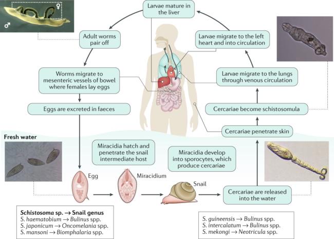
For the unfamiliar, S. haematobium is the parasite behind schistosomiasis — also called snail fever, or bilharzia. Its eggs are normally only encountered in clinical samples from infected patients, which makes them critical for diagnostic development yet difficult to obtain in a controlled laboratory setting. Our vial has since been divided into aliquots bound for two research hubs: the Prakash Lab at Stanford’s Shriram Center for Bioengineering, and Dr. Lee’s lab at UTHealth in Houston.
From a many-people-CC’d email thread to a (totally-not) high-security handoff of a globally significant pathogen, this small episode is what frugal science really runs on: serendipity, community, and scientific generosity.
A friend who connected the dots
Maya Grayck — my dear friend and coworker in Clyde Wright’s lab at CU Anschutz from 2021 to 2023 — joined our team and this project in March. We’re glad to have her for many reasons, but one stands out: without her knack for connecting dots across states, institutions, and labs, we simply would not have access to these samples.
In March, Maya got up to speed on a Zoom call with Jim Cybulski, Manu Prakash, and the Health in Your Hands team — and within a few days she realized our worlds overlapped in an unexpected way. Her mother, Dr. Eva Nozik — mentor and friend to so many — had just studied pulmonary hypertension from chronic schistosomiasis in a murine model, alongside Brian Graham, who keeps schisto specimens in his lab at UCSF.
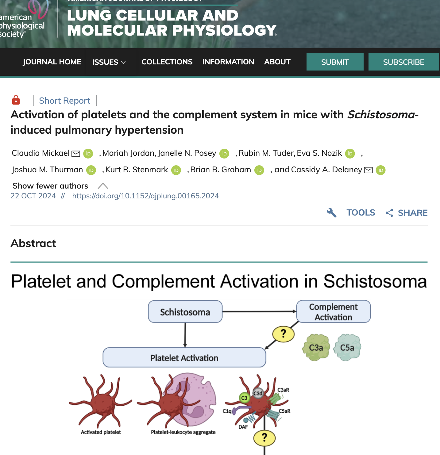
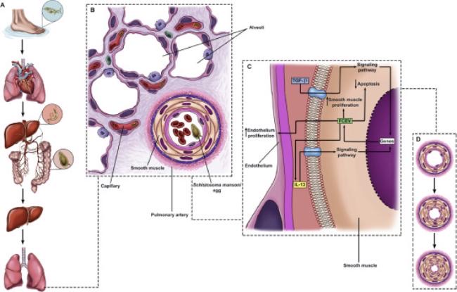
Eva is the kind of physician-scientist who studies — at the highest level — just about every nuance of an organ system, so I should have known we’d be seeking her sage advice eventually, given schistosomiasis can reach the lungs: her area of expertise. At that point, I was still naive to that clinical consequence.
Why these eggs, specifically?
A fair question we kept getting: why not just use the eggs already in Dr. Lee’s lab — aren’t those good enough? Both S. mansoni and S. haematobium are parasitic flatworms that cause schistosomiasis, but they differ in where they settle in the body, what their eggs look like, and how the disease presents.
S. mansoni
- Target
- Intestinal tract & liver
- Eggs in
- Stool
- Presents
- Intestinal & hepatic disease
S. haematobium
- Target
- Urogenital system
- Eggs in
- Urine
- Presents
- Hematuria; long-term bladder damage & cancer risk
This distinction is critical for diagnostic development. Urine-based diagnostics — especially the low-cost, field-deployable kind we’re building — must be tested using S. haematobium eggs specifically, since they differ in size, shape, and localization from S. mansoni eggs.
A tiny rented car, a big errand
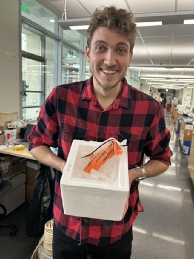
In early April, Maya joined myself, Phillip, and Nehali on a trip to Stanford. Anesta Kothari and Hope Leng — who are working on a low-cost molecular test for schistosomiasis — coached us on how to ask for the eggs and how to transport them. We opted for frozen samples on dry ice.
A couple of days in — realizing time was running short — I asked Phillip and Maya whether they’d take our tiny rented car and run the mission to UCSF without me, while I stayed on campus, another coffee in hand, prototyping and testing the microfluidic trap we were developing. They accepted the mission.
What we’ll do with them
Here’s the mile-high view of what our Stanford collaborators and friends plan to use the samples for — three reasons these specific eggs matter to the work ahead.
Why we want these eggs in the lab
Develop a workable field diagnostic
Prove out a urine-based method a community health worker can run with a Foldscope — far from any formal lab.
Build a sustainable workflow
Move from one-off samples toward a repeatable, affordable sample-prep and testing pipeline that holds up in the field.
Test Vaibhav's PlanktoScope
Run these samples through an automated imaging setup to help train its model. See the project →
We’ve joined forces with Vaibhav Shokeen for the current study, and we’re thrilled to be meeting in five weeks in Nigeria. In the next lab note, you’ll see how we use these samples to help train his automated model. More to come — stay tuned, my friends.
You’ve launched us into 40%-funded territory.
Thank you for seeing the vision — a future with accessible diagnostic solutions to this neglected tropical disease. Please feel free to share these lab notes with any friends, family, or colleagues who’d want a look behind the scenes of what this work takes.