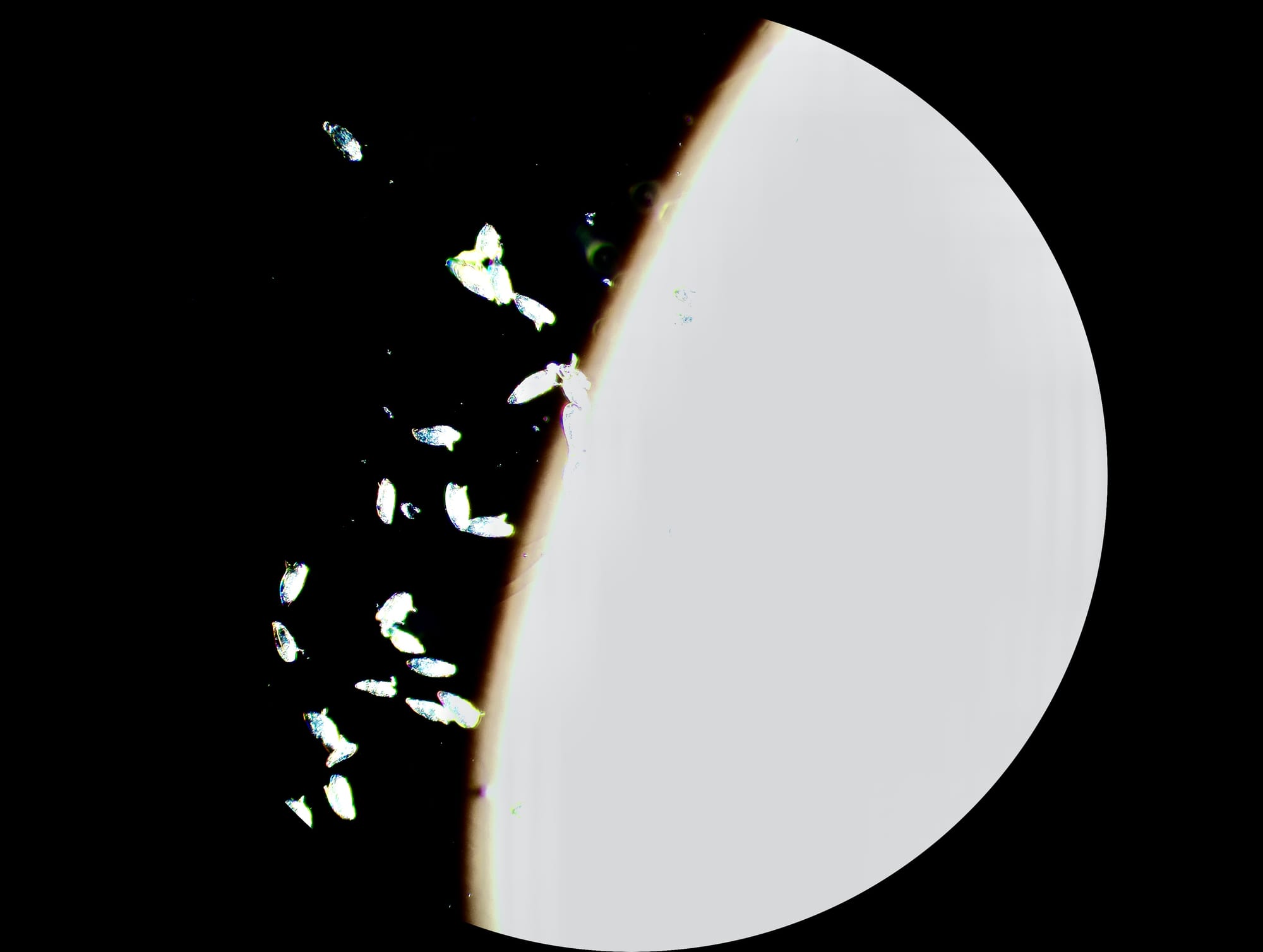
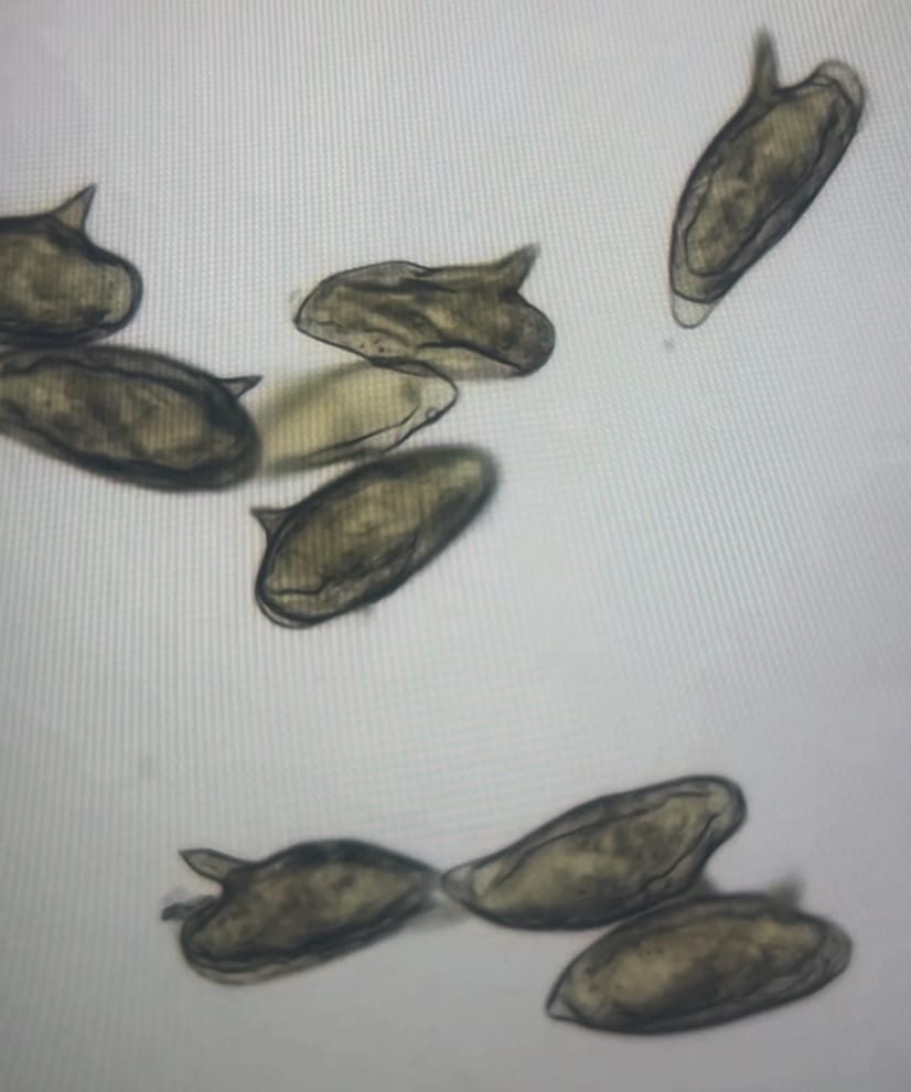
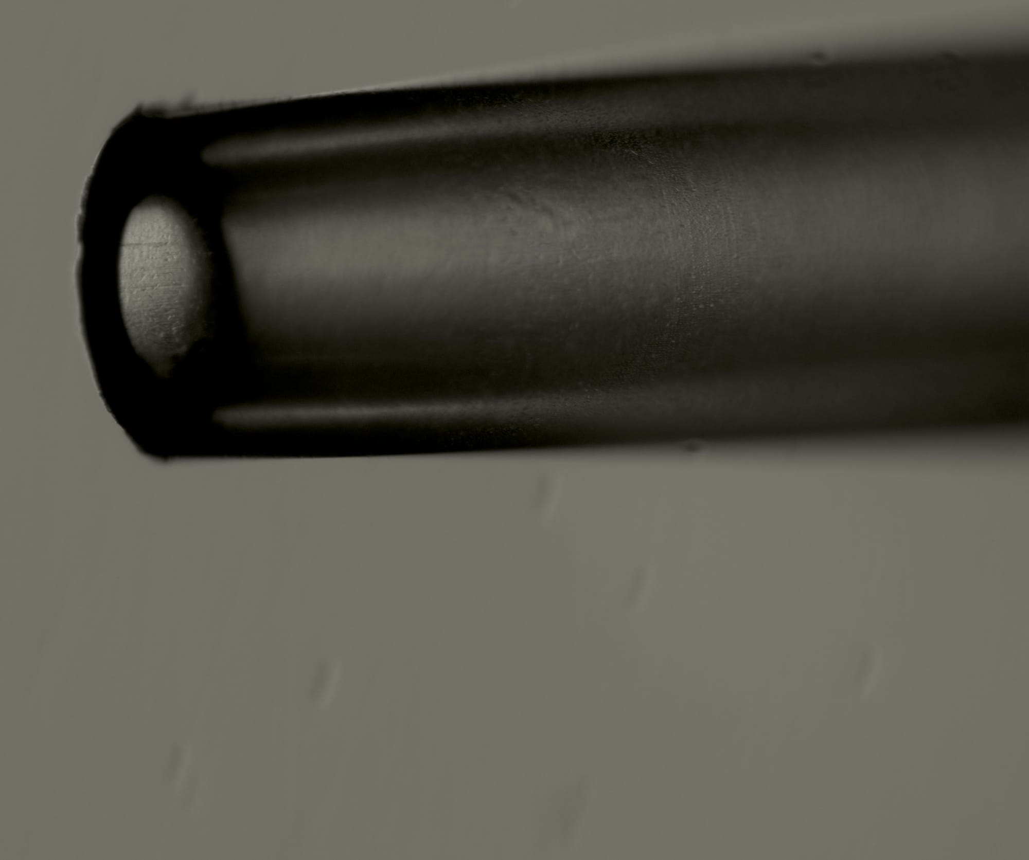
 Underlit eggs in a water droplet
We’re trying to build a mobile, handheld lab for diagnosing schistosomiasis in a community-led way, one that could let community health workers help control the disease sustainably and affordably. This month, our prototype started doing the thing.
The goal is simple to say: filter parasite eggs out of a urine sample so they can be viewed under a microscope, with a toolkit that’s field-friendly and reusable. Schistosomiasis is a neglected tropical disease that leads to serious complications like bladder cancer, and it mostly affects people who rely on natural freshwater in sub-Saharan Africa. Here are two experiments, two weeks apart.
Can we count what we catch?
I tried the SchistoFilter v2 (at the time, a slanted-wedge trap newly redesigned by Dr. Cybulski) on S. mansoni eggs suspended in a small volume of water. The device wasn’t fully assembled (only two of the corner tighteners were securely attached); my main goal was a quantitative approach. Can we count the eggs in a water droplet under the microscope, suspend them, filter them, and count how many the device caught?
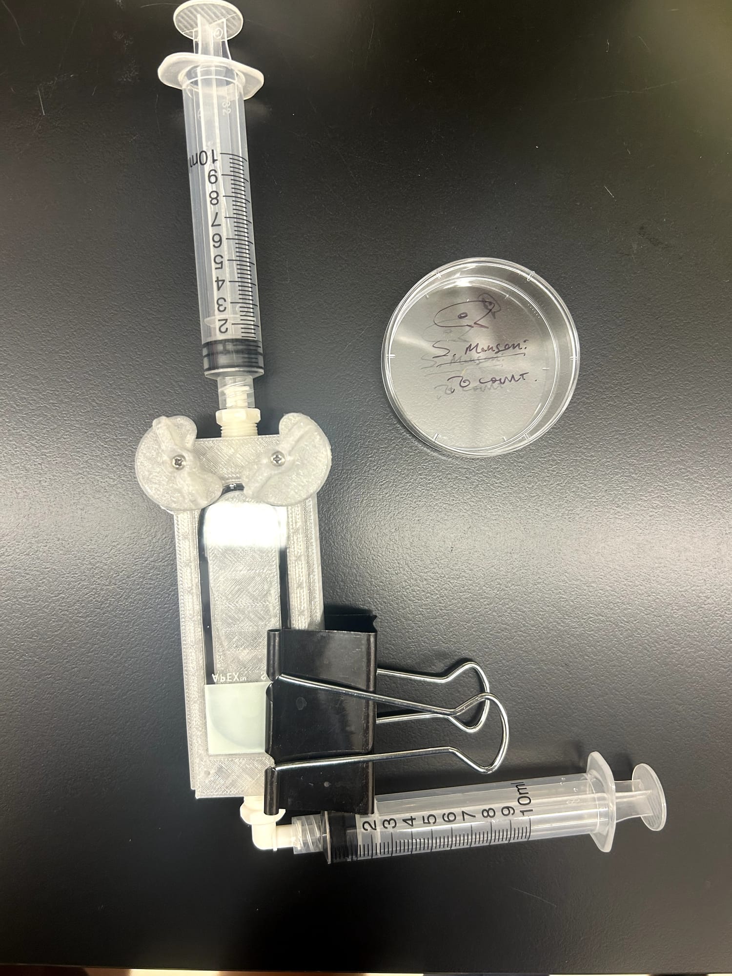
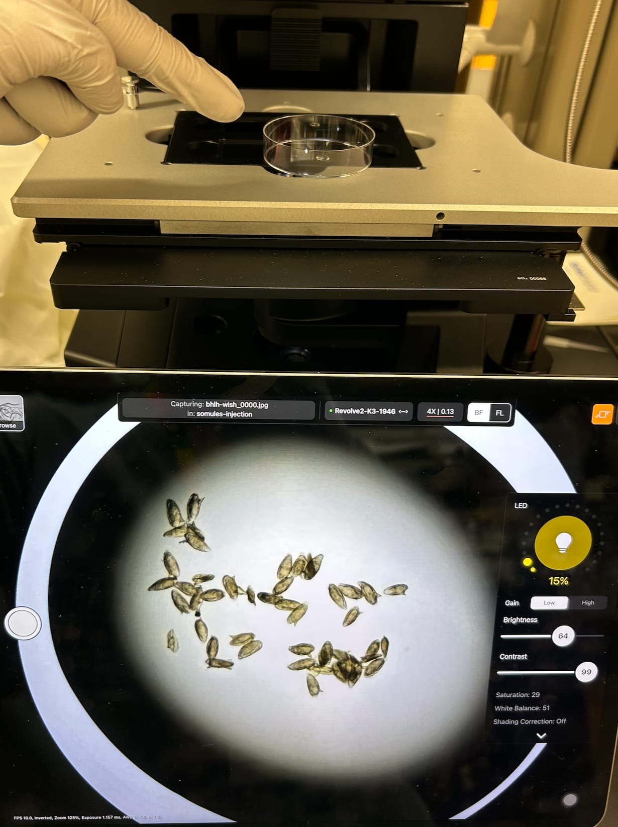
I hypothesized that the device would catch 25–50% of the eggs. The results surprised me: it trapped about 75–80%, roughly 37 to 42 of 50 eggs. I was also curious how long cleaning would take between uses: you can disassemble, rinse, and reassemble the device in 1 minute and 40 seconds, with some fumbling and fidgeting.
All the visible eggs sat within a thin strip of the wedge — its “landing strip.” That’s encouraging: it suggests the device works as intended, a slanted, size-based filter that eggs can’t pass through but urine and white blood cells can, on their way to the collection syringe.
Thank you to Anesta Kothari, who had the idea of pipetting the eggs and counting them under the microscope. It lets us keep testing how well each version performs.
What we learned for the next version
Leaks
Fluid leaked at the inflow and outflow tracts; a seal was only possible with extra adhesive and a binder clip.
Hardware
Four twist-lock corners instead of a binder clip, and better screws for the corner clamps.
Field cleaning
Caroline pointed out that pressurized tap water won’t be available in rural clinics, so we should test cleaning with poured fluid.
Eighty-five percent
Two weeks later: the v5 prototype, with Dr. Cybulski’s freshly printed v2 corner-securing twist pieces, and the in- and out-flow tracts secured with parafilm. We hypothesized that from 64 counted S. mansoni eggs, we could filter more than two-thirds and see them under the microscope. We counted 55 of 64 — 85% trapping efficiency.
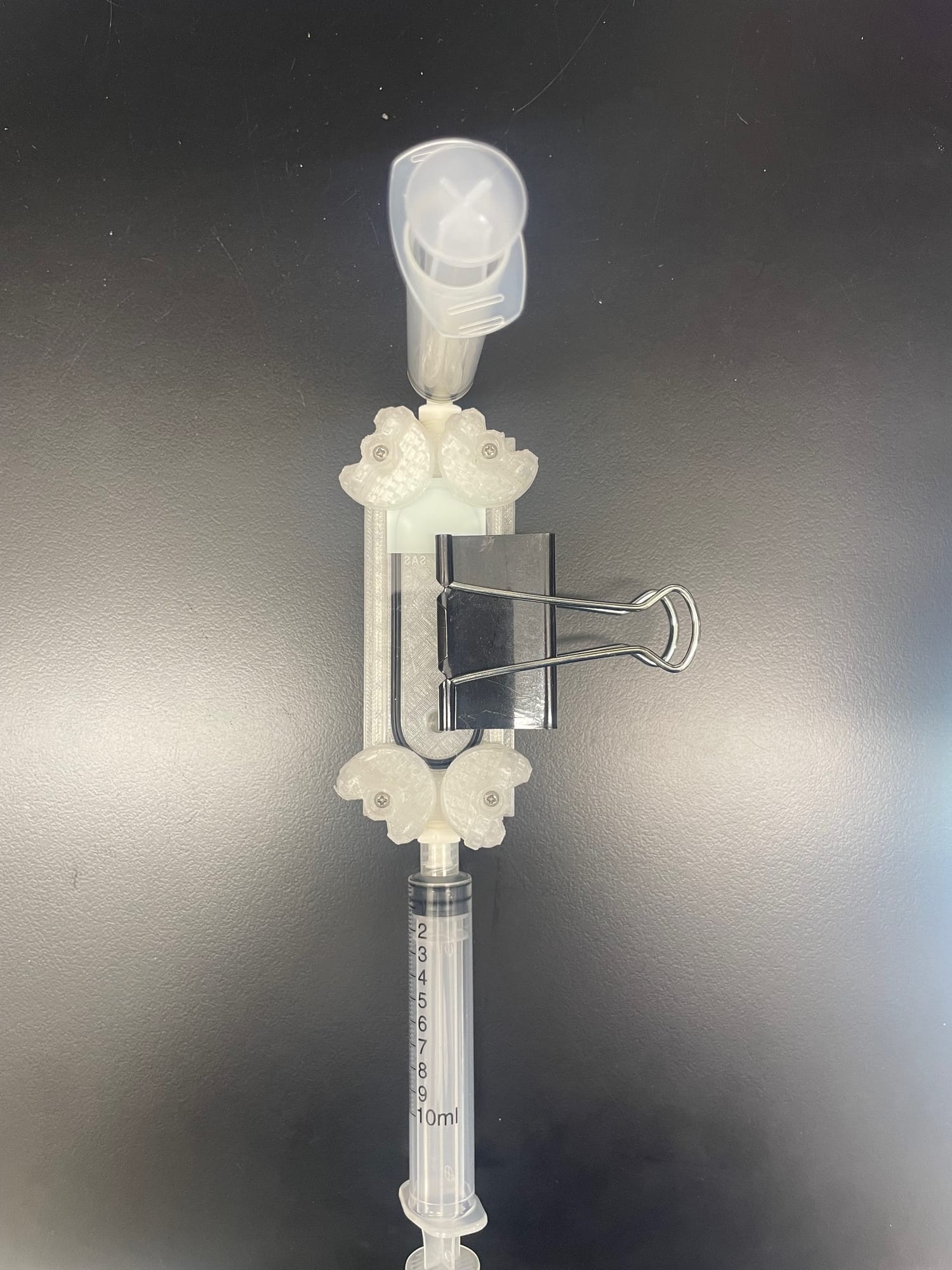
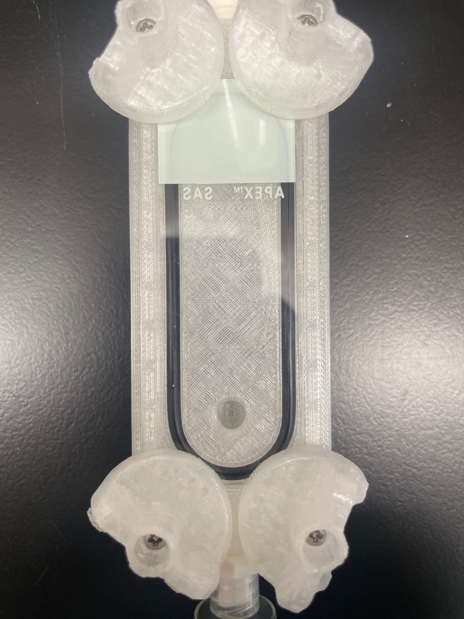
The key “aha” wasn’t the number. It was realizing we’d built a workflow — for testing the device, sharing results, and spotting what to improve next.
The counting workflow
Count
Painstakingly count the eggs in a single 10 µL drop on a petri dish.
Dilute
Pipette the drop into a tube and dilute to a 5 mL sample.
Check the tip
Look at the pipette tip under the microscope for stragglers, and account for any (this time, none).
Filter
Draw the sample through until you feel the “tug” Jim coached us to look for, the sign of a sealed system.
Recount
Count the eggs trapped in the device.
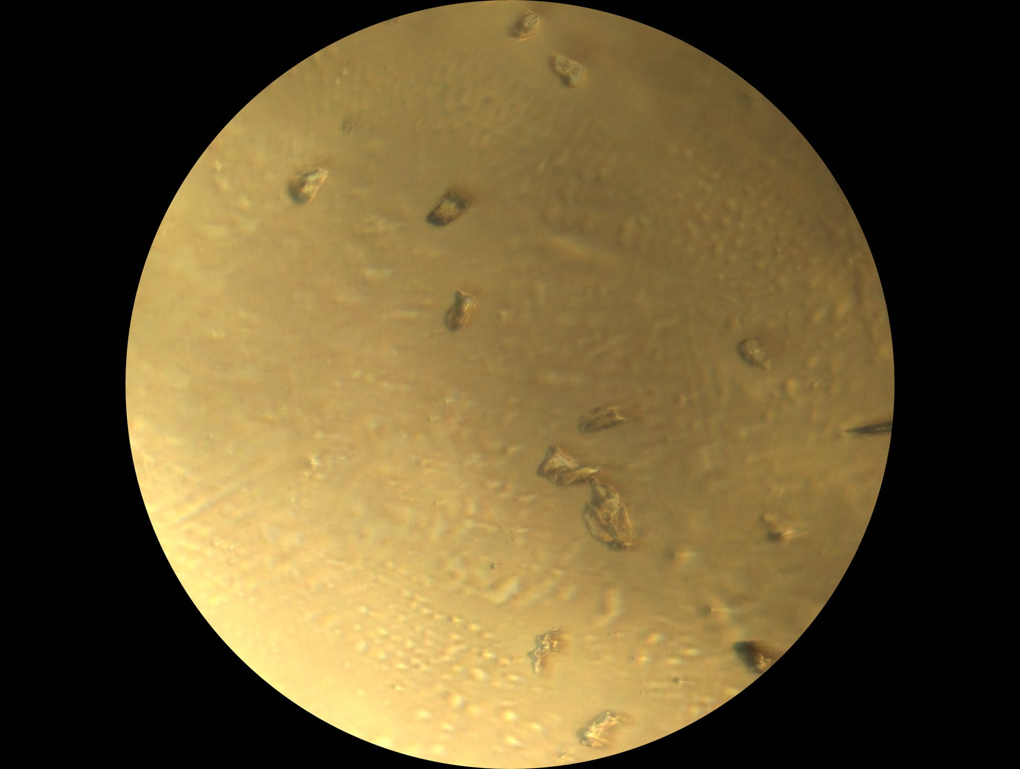
Most intact eggs were trapped earlier than expected (about a third of the way up the staircase), while smaller fragments caught further along in the narrower channels. The leaks aren’t solved, and we’d like to secure the slide without extra hardware. Jim has already sent updated files for v6, which we’ve printed; we’re just waiting on new Luer fittings from McMaster.
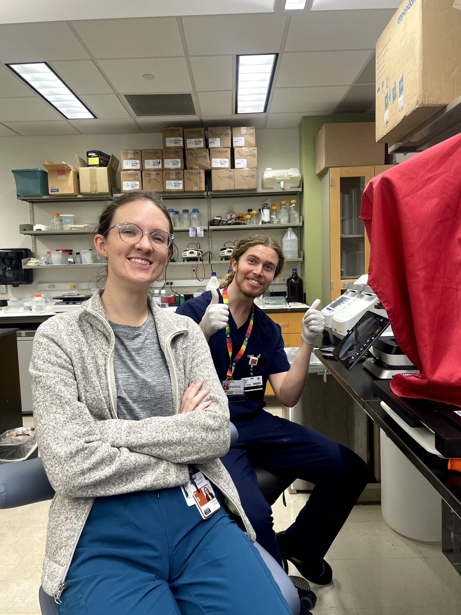
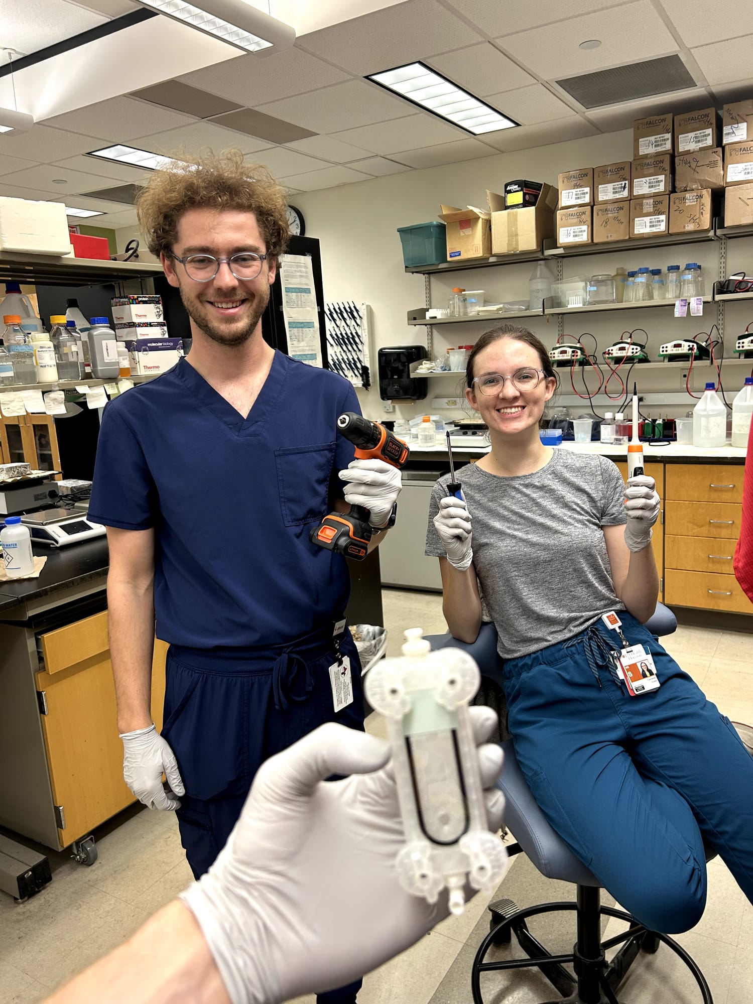
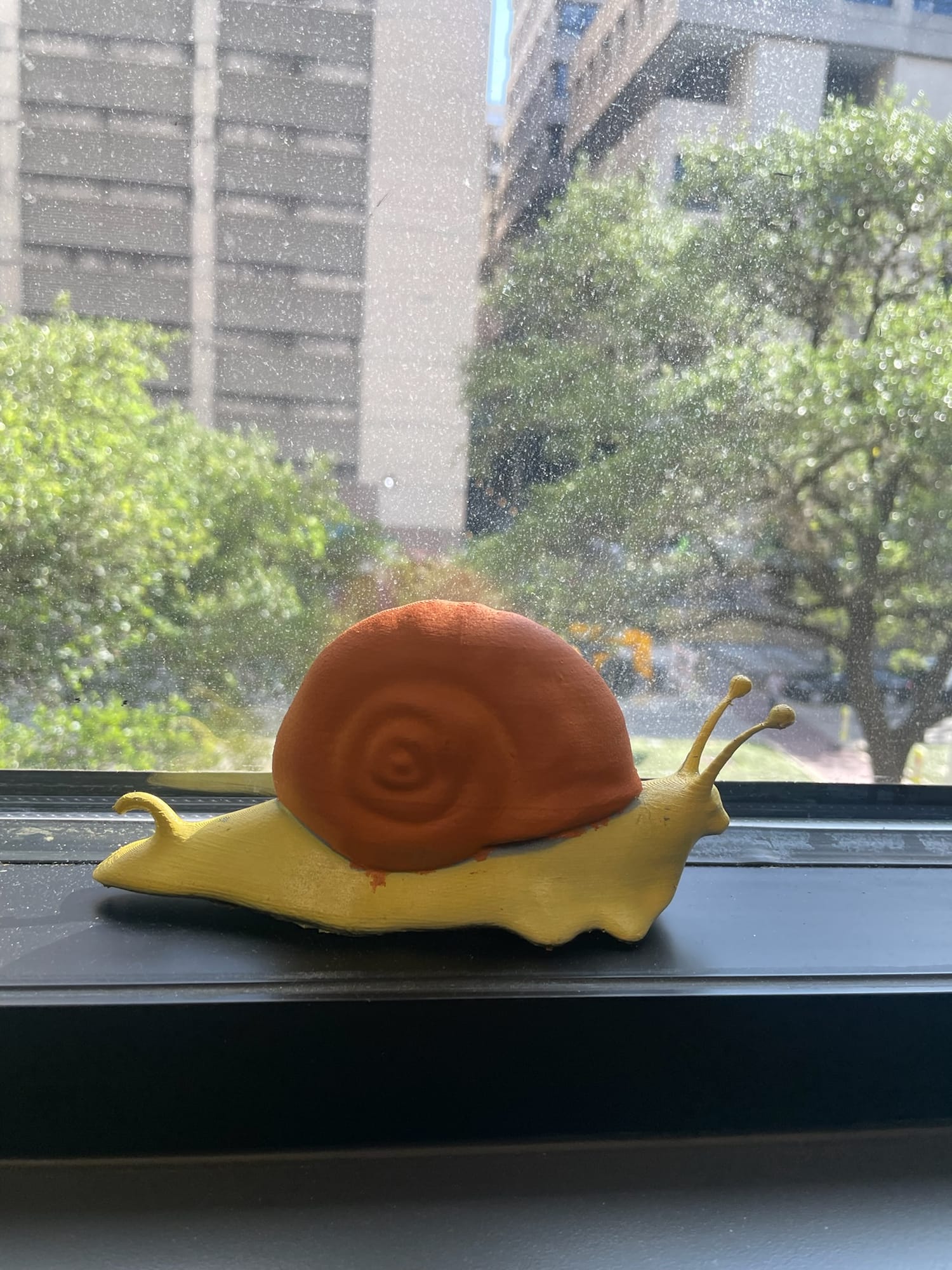
Thank you to Jon, who found the right screws; Phil, who printed the new pieces; Simon and Caroline, who helped count and filter eggs in Dr. Lee’s lab; and Sabona Simbassa, whose coaching on the microscope made all of it possible. To thank the Lee Lab for putting up with our experimentation shenanigans, we gave them a hand-painted, 3D-printed snail (thank you, Phil, Morgan, and Jon).
One count at a time
Every version teaches us something the last one couldn’t. Thank you for funding the iterations.